
This week in science history: The x-ray was first exhibited on January 18th, 1896.
If you’ve ever broken or fractured a bone, you’ve probably had an x-ray taken! These machines, invented by German physicist Wilhelm Roentgen, allow doctors to see inside your body to better understand a variety of health problems.X-rays are similar to visual light, both are forms of electromagnetic light. X-rays, however, have considerably more energy than visual light and can pass through most objects. This is what allows x-ray technicians to see inside your body!
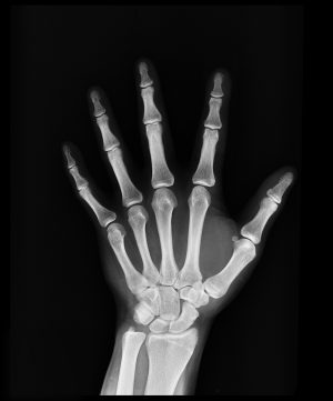
Images produced using x-ray machines are called radiographs. X-ray technicians use radiographs to look at the tissues and structures inside your body. When you have a broken bone, your doctor is able to look at the radiograph and see exactly where break is and how bad it is. This information is helpful in deciding how to best treat the injury.
Other uses for x-rays include:
- they have been used to examine old paintings to see if there is anything underneath the artwork
- they have been used to study ancient fossils
- they have even been launched into space!